Brain tumor detection using machine learning that includes the application of image processing and deep learning methods. For all levels of students, we share best ideas and topics on their field. Originality is assured as it is our main work ethics. Scientific and experimental methods based on practical solutions will be shared. Here, we deliver the prominent sketch of a brain tumor detection project:
- Objective :
- We identify the existence of a brain tumor with the help of Magnetic Resonance Imaging (MRI) scans.
- Dataset :
- From the Kaggle platform, the publicly available datasets like Brain Tumor Dataset or Brain Tumor Segmentation (BRATS) datasets used by us that are supplied through MICCAI challenge.
- Project Steps:
- Data Preprocessing :
- Image Resizing: We resize all the images to attain similar dimensions.
- Normalization: The pixel values are standardized within the value between 0 and 1.
- Data Augmentation: The dataset is extended with rotations, flips, zooms and more for raising the strength of our model.
- Feature Extraction:
In the conditions of deep learning models like Convolutional Neural Networks (CNNs), the feature extraction is naturally performed by the model itself. Yet, we utilize the usual methods that includes,
- Segmentation: The area of interest is separated from the background.
- Texture Analysis: Texture based features are derived by us that possibly reveal the existence of tumors.
- Model Building:
- Traditional Machine Learning Approach: Make use of algorithms such as SVM (Support Vector Machine) or Random Forest associated with hand-crafted attributes.
- Deep Learning Approach: We execute a Convolutional Neural Network (CNN) that mechanically understands the hierarchical characteristics from the MRI scans.
- Transfer Learning: The pre-trained models deployed in this like VGG16, ResNet, etc. and manage them for performing tumor detection tasks.
- Model Training :
- The data is categorized into training, validation and test sets.
- The selected model is trained by us on the training dataset while the performance of our model is examined in the validation set.
- If it is required, regulate the hyperparameters.
- Model Evaluation:
- Approach the evaluation metrics appropriately,
- Accuracy: It depicts the percentage of categorized images accurately.
- Precision, Recall, and F1 Score: If class imbalance occurs, it is very essential for balancing the datasets.
- ROC Curve and AUC: This is beneficial for us in performing binary classification problems.
- On the test dataset, we test the model to estimate the performance.
- Post-processing:
Some post-processing methods are valuable for deep learning models. It includes,
- Morphological Operations: Dilation and erosion operation is employed by us in enhancing the tumor limitations in the isolated image.
- Thresholding: The prospected map which is produced from the model is transformed into a binary mask signifying the tumor affected regions.
- Visualization:
The results are figure-out that distributes clarity about model’s decisions:
- Heatmaps: Our model detects the tumor affected areas in the MRI scans.
- Overlay: The detected tumor regions are superimposed on the original MRI to figure out the appropriate location and size.
- Deployment (optional):
- While we are satisfied with model performance, then approach our method in a mobile application or webby by utilizing platforms such as Flask, Django, or TensorFlow Lite for on-device assumption.
- Methods and References:
- Image Processing: OpenCV and Scikit-image are the methods involved in this process.
- Machine Learning: Scikit-learn is particularly included in ML.
- Deep Learning: Deep learning consist of tools like TensorFlow, Keras and PyTorch.
- Deployment: We approach models such as Flask, Django, TensorFlow Serving and TensorFlow Lite.
- Obstacles and Reflections:
- Class Imbalance: Even if there are some images that appear with tumors or few not, it is required for reporting during the training period of the model.
- Data Variability: Depends on the utilized machine, MRI scans are varied like the patient’s position, etc. Our model must be strong to tackle this type of variation.
- Evaluation Metrics: Apart, accuracy is not the best metric especially in case, if the data is not balanced.
Dive deeper into the project that is elaborated into tumor subtypes, levels, and other features for supplying an extensive diagnostic tool. Make sure that our created tool is just approached as an add-on tool for the diagnostic process; it does not replace the instructions of clinical experts in validation.
Brain Tumour Detection Using Machine Learning Project Thesis Ideas
Get customized thesis solutions from phdtopic.com professional team. All the simulation results will be explained we follow the protocols as per your norms. Article writing, along with research proposal and topic assistance much more are provided by us.
- Efficient Brain Tumor Detection Method Using Feature Optimization and Machine Learning Algorithm
Keywords
MRI, Brain tumor, Machine learning, SVM, DWT, Firefly
To identify the tumors, we recommended a multi-stage support vector machine technique in our research. This technique increases the area of coverage across overall MRI image features. We performed feature selection and optimization process by employing firefly method. Discrete wavelet transform is utilized for feature processing. We have done a comparative analysis for our suggested method with other current methods like SVM and CNN.
- Detection of Brain Tumor using Machine Learning Classifiers – A Comparative Study
Keywords
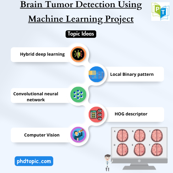
Deep learning, Hybrid deep learning
Several ML methods like DT, Discriminant Model, KNN, Ensemble, SVM, NB, Neural Network and Kernel are compared in our project to identify the best method for the detection of brain tumor. We performed segmentation procedure to acquire the specific region of interest from the MRI images. Then we extracted the features from the segmented region by utilizing GLCM technique. Here we executed an integrated DL approach to detect the brain tumor.
- Detection of Brain Tumors through the Application of Deep Learning and Machine Learning Models
Keyword
Convolutional neural network
To display the variety of MRI images, a high-pass filter is utilized in our approach. A high median filter is utilized to integrate with slices. We preprocessed the MRI images by smoothing process and the borders are highlighted to enhance the quality of images. We employed various supervised ML methods like SVM, KNN, RF, NB, and CNN to train the model. As a result, CNN method outperformed other methods.
- Machine Learning based Brain Tumor Detection and Classification using HOG Feature Descriptor
Keywords
Computer Vision, HOG descriptor
An innovative approach is suggested in our paper to easily identify and categorize the brain tumor thorough the utilization of MRI. We preprocessed the data to remove the noise. Then we extracted the relevant features by using HOG feature descriptor. To examine the accuracy of this approach, several ML methods such as SVM, KNN, Gradient Boost, XGBoost and LR are employed. Results show that, XGBoost method offers better end results.
- An automated brain tumor detection and classification from MRI images using machine learning techniques with IoT
Keywords
Level–level set, GLCM, KNN, Chan-vese
To detect and categorize the brain MRI pictures as tumor and non-tumor areas, different ML approaches are employed in our project. We preprocessed the images to eliminate the noise by using median filtering. After that, we segmented the image by employing Chan-Vese method. A GLCM is utilized to extract the tumor related features. From that, the essential features are selected. We executed two-class classifier by utilizing SVM and it is verified with KNN method.
- Exploring the Effectiveness of Various Machine Learning Algorithms for Detecting Brain Tumors in MRI Images
Keywords
VGG 16, VGG 19, DenseNet121, Resnet50, LeNet
A main objective of our study is to identify brain tumors through the utilization of MRI. We employed several techniques such as artificial neural network (ANN), convolutional neural network (CNN), VGG 16, VGG19, DenseNet121, Resnet50, and LeNet in the brain tumor identification process. As a consequence, VGG-19 technique provides greater efficiency when compared to other methods.
- Brain Tumor Detection using Machine Learning and Convolutional Neural Network
Keywords
Brain MRI tumor images, Otsu Thresholding, Local Binary pattern
A binary classifier is constructed in our suggested research to identify brain tumors by utilizing MRI pictures. The ML based approach and convolutional neural network method is employed in our work for the MRI related brain tumor identification. MRI images with tumors and non-tumors are considered for detection process. We conclude that, when compared with ML techniques, CNN method provides greater results.
- Morphogenesis Method to Detect Brain Tumor using Machine Learning Technique and Noise Filtering
Keywords
Anisotropic filtering Brain Tumor, Morphogenesis
In our project, brain MRI pictures are examined to detect the brain tumor. We eliminated the noise exists in the images by utilizing anisotropic filtering. Then we segmented the image by employing SVM. To extract the infected region, feature extraction process is carried out. By discovering the frequencies of the pixels exist in the noise eliminated MRI picture, we detected the tumor affected region. As a result, Genetic Algorithm and SVM offer better performance.
- Features Driven Brain Tumor Detection Using Machine Learning Models
Keywords
Brain tumor detection, Classification, BraTS 2015, Random Forest
A feature driven brain tumor identification framework is recommended in our study. We preprocessed the images and it is transformed from .nii.gz format to NumPy arrays and it is split into various subsets. To remove the unessential background, we cropped the images. Then we carried out the feature extraction process and GLCM generation. To train the framework, several methods like SVM, RF and KNN are employed. In that, RF outperforms other methods.
- Execution Analysis of Machine Learning Technique Based Detection and Classification of Brain Tumor from MRI images
Keyword
GLCM Brain tumor
Various ML techniques are discussed in our article to differentiate and classify the image as tumor and non-tumor region. We extracted the relevant features from the cancerous area by using gray level co-event network method. After that, we selected the important features. We employed SVM method to perform two-class classifier and it is compared with KNN technique. We states that, our suggested model obtains greater results than other existing methods.