Skull stripping is the process of skull removal, which is used in the process of brain tumor segmentation. The medical images of the brain offer information about the internal structure and thus it can aid in the detection of a variety of conditions, including malignant glioma. The intensity of the tumor and the skull are similar, making automatic tumor diagnosis challenging. Like the pre-processing stage for recognizing the brain tumor, an algorithm for skull stripping sought to address such an issue.
Through this article, we provide you with a complete picture of skull stripping brain tumor segmentation which will be very much essential for your research project in the field.
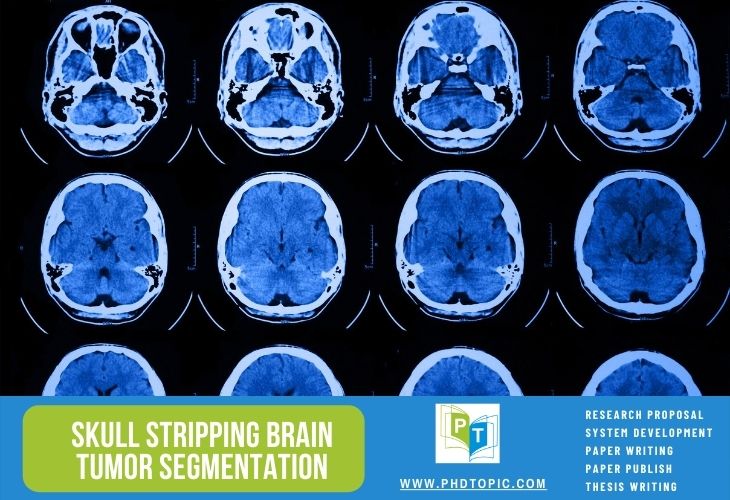
Overview of Skull Stripping
An adequate skull stripping is necessary due to the complicated morphological structure of the brain and intensity effects on brain MRI Images. Skull stripping refers to a procedure that involves removing a portion of the human skulls for clinical study in segmentation of brain assignments, and its precision and effectiveness are critical for medical testing. Let us first start by looking into the constraints involved in skull stripping,
Limitations of skull stripping
- It is very hard to separate the required brain tissues of the other tissues and determine the efficient size of morphology
- Even very small variations can lead to huge differences does every aspect of imaging is very sensitive
- The threshold value at the initial stage determines the performance of segmentation
- For images with inhomogeneous intensity, the result is obtained with reduced or over-segmentation
- Also the variations in intensity affect over-segmentation and under segmentation of the brain images
- When intervened by artifacts, better skull stripping images cannot be obtained
- Whenever the brain images have comparable intensities profile towards the non-brain imaging, the morphological operation might fail to differentiate between the brain and non-brain aspects.
- It can’t be used on the abnormal brain (tumors) since the atlas-based methodology needs anatomy, but tumors can change the brain’s structure considerably.
The effectiveness of diagnosing tumors, pre-surgical preparation, cortex surface reconstruction, and cerebral morphometry is influenced by the precision of the skull stripping procedure, which is regarded as an important stage in brain tumor segmentation. The cranium is removed to decrease the possibility of mislabeling sick components. The technique of skull stripping is extremely problematic due to the following complexities
- Human brain complexity
- MR scanner parameter variations
- Unique features
- Processing images with less contrast and quality
To decrease these impacts, several strong skull stripping algorithms have now been developed. Our technical experts have gained huge expertise in skull stripping brain tumor segmentation algorithms and by providing proper real-time implemented examples we will also answer all your concept-based queries if any. What are the models available for skull stripping brain tumor detection and segmentation?
Skull stripping methods for brain tumor segmentation
stripping methods for brain tumor segmentation
- Morphological functions and histogram-based analysis and thresholding
- Threshold methods, seed growth, and morphological processing
- Two-dimensional region growing and three-dimensional morphological operation methods
- Connected component analysis and anisotropic diffusion filtering
- Diffusion and two-dimensional Marr Hildreth operator based edge detection
- Bridge burner method for narrow connectivity removal and intensity threshold
To get research-related facts and authentic quality numerical data on all these methods you can get in touch with our experts. We are here to supply you with proper guidelines, support, and in-depth Research guidance needed to carry out advanced skull stripping tumor segmentation projects. Let us now talk about the description of brain images
Brain images description for skull stripping
The following is a description of the methods in which different input images are distinguished
- T1 is used to refer the T1-weighted MRI images
- T2 refers the T2-weighted MRI scans
- T1c stands for T1-weighted MRI by enhancing the contrasts
- T2 – Flair stands for T2-weighted MRI with fluid attenuated recovery of inversion
The images are 512 x 512 pixels in size. Repetition Time (or TR) and Echo Time (or ET) are the two parameters that affect tissue contrast (or TE). A paramagnetic contrast medium (gadolinium) is used in T1 contrast augmented imaging to decrease the T1 relaxation time and boost the signal strength, allowing for a sharper view of the areas damaged by bleeding. Here is a quick note on how the different parts of the brain are indicated in a scan
- White matter
- T1 – Weighted, T2 – weighted, Flair and T1c are respectively represented in light, dark gray, dark gray and light
- Inflammatory findings
- T1 – Weighted and T1c are respectively represented in dark and dark gray while T2 – weighted and Flair are represented as bright parts
- Fat within the bone marrow
- T1 – Weighted, T1c and T2 – Weighted, Flair are respectively represented in bright and light colours
- Cerebrospinal fluids
- T1 – Weighted, T2 – weighted, Flair and T1c are respectively represented in dark, bright, gray and dark gray
- Cortex
- T1 – Weighted and T1c are respectively represented in gray and dark while T2 – weighted and Flair are represented as light gray
In addition to these representations, you are also required to have deep insight into the imaging methods, techniques, approaches, and algorithms for Skull stripping brain tumor segmentation. For this purpose, you can readily reach out to us. With the help of our world-class certified engineers, developers, and experts we are offering one of the most reliable and professional research guidance in the world. Let us now look into the stages involved in skull stripping
What are the steps in skull stripping for brain tumor segmentation?
- Inputs
- T1 – weighted MRI image is given as input. It is considered to be the most optimal image sequence for brain tumor analysis
- Image Normalization
- Pixel value intensity is altered for performing input MRI image normalisation
- Image Registration
- The MRI image given as input is then registered where data from multiple sets are converted into single system of coordinates
- Skull stripping
- The algorithms like differential revolutionary and artificial bee colony algorithm are used in removing non cerebral tissues and skull images
- Removal of noise
- Noise removal is performed using anisotropic diffusion filter the process which is considered to be the most important for brain tumor detection
- Segmentation
- MRI image segmentation is the last and final step
We are here to help you design better projects for skull stripping by incorporating advanced methodologies and working with all kinds of tools, libraries, and approaches associated with these stages. Check out our website for the successful projects that we have completed. We will now talk about the skull stripping brain tumor toolboxes
Tools and toolboxes for skull stripping brain tumor segmentation
- Python
- Several libraries, such as skull stripping, skull anatomy, and so on, are available in Python. Choose Deep Learning based incoming brain image processing technologies with an emphasis on sensitivity and precision with the deep brain library.
- The accuracy of the observations is measured by analyzing the output to the existing Analyze Skeletons Fiji plugin and an innovative scanning electron microscopy (SEM)-based approach.
- Python data structures like SciPy sparse matrices and pandas data frames additionally allow the findings to be analyzed inside the Python ecosystem of scientific libraries.
- MATLAB
- We begin by choosing input image data from the retrieved dataset.
- Regarding image processing, analytics, visualization, and algorithm development, MATLAB provides for the image processing Toolbox, which includes a full set of reference-standard techniques and workflow approaches.
- Picture segmentation, image enhancement, noise removal, geometric transformations, registration of images, and three-dimensional image processing are also available.
- It can read and write a variety of specialized picture file types.
- Scilab
- Upload the dataset first, then choose the input test images to be analyzed.
- The SCILAB framework and implementation of Blocks, skeletal architecture that would be immediately installed at SCILAB launch, or programs with Graphical User Interface are used to construct the skull identification system which is based on picture pre-processing, image segmentation, and ultimately identifying the skull match procedures.
- OpenCV
- Construct a skull identification scheme that accepts binary images or BLOB files as inputs.
- Then use the skeletonization technique to reduce foreground portions in a binary source image to a skeletal remainder that retains the majority of the initial region’s dimensions and connection.
- The imutils skeletonize program can then be used to create the topology aspects of the skeleton of the images.
- Following that, you can apply additional processes such as object selection, extraction, modification, comparison, registering, tracking, recognizing, compressing, and so on.
Usually, our technical team provides you with all the essential information in the form of theoretical and practical demonstrations that you will also be allowed to interact with potential, renowned, and top researchers from around the world with whom we have established alliances. Therefore by organizing and planning your research work we assure you that you have an enjoyable career. Let us now look into some of the important databases for skull stripping
Datasets and databases for skull stripping
- OASIS-3 Clinical, (Longitudinal Neuroimaging, and Cognitive Dataset for Alzheimer’s Disease)
- It seems to be a retrospectively accessible data compilation for the above thousand subjects gathered over three decades by the WUSTL Knight ADRC through multiple current initiatives.
- Respondents’ ages range from forty-two to ninety-five years old and comprise about six hundred cognitively normal adults and more than four hundred and eighty people in varying levels of cognitive deterioration.
- Most members were provided a randomly generated identity, as well as all times, were deleted and normalized to represent the number of days since they first entered the research.
- FLAIR, T1w, ASL, T2w, and SWI, duration of travel, resting-state BOLD, and DTI sequences are among the nearly two thousand MR trials in the collection.
- Several of the MR Sessions include segmentation files generated by Freesurfer processing.
- PET imaging comprising three separate tracers, PIB, AV45, and FDG, including above one thousand five hundred original imaging data, as well as the associated post-processed records from the Pet Unified Pipeline (PUP), are indeed included in OASIS-3.
- OASIS-2 (Longitudinal MRI Data in Nondemented and Demented Older Adults)
- It refers to the longitudinal study of about one hundred and fifty people aged between sixty and ninety-six years
- For a maximum of about three hundred and seventy imaging examinations, each individual has been evaluated on two or even more periods spaced by at a minimum of one year.
- Three or four separate T1-weighted MRI images taken in single scanning sessions are presented for every participant.
- Every participant is right-handed, and both males and females are included.
- All through the trial, about seventy-two of the participants were classified as non-mentally ill.
- Nearly sixty-four of the participants, comprising fifty-one people with minor to moderate Alzheimer’s disease, were classified as dementia patients at the moment of their first assessments and maintained so for successive examinations.
- Additional fourteen participants were classified as non-dementia at the point of their initial evaluation before being reclassified as demented at a future appointment.
- OASIS-1 (Cross-sectional MRI Data in Young, Middle Aged, Nondemented and Demented Older Adults)
- The cross-sectional sampling of four hundred participants ranging in age from eighteen to ninety is included in this dataset.
- Three or four separate T1-weighted MRI scans taken in single scanning sessions are presented for every individual.
- All of the participants are right-handed people, and then both women and men are included.
- Well over the age of sixty, about one hundred of the participants were medically diagnosed with really weak to intermediate Alzheimer’s disease (AD).
- A dependability dataset is also supplied, which contains about twenty nondemented participants who were examined again in about ninety days of their initial orientation phase.
Our experts have gained a lot of expertise in handling these databases as we have delivered several projects in skull stripping using them. Also, we are here to update you regularly on all the recent advancements and we ensure that your potential novel ideas will be brought into reality. Hence we are here to support you holistically throughout your research. Let us now talk more about the parameters used for analyzing the performance of skull stripping brain tumor segmentation systems.

Performance metrics for skull stripping
The following are the important parameters of the metrics used in analysing the performance of skull stripping methods
- False positive and false negative rates
- Dice similarity coefficient or DSC
- Jaccard similarity coefficient or JSC
DSC and JSC are used in determining the similarity between real and skull stripping images. You can find a detailed description of all these parameters and performance metrics of Skull stripping brain tumor segmentation from our website. We have been offering customized desert support to students and Research scholars from all around the world. So you can contact our experts with greater confidence for ultimate research support in skull stripping.